
The causes of back pain are often multi-factorial, which can make the process of back pain diagnosis extremely complex. While some diagnoses of back pain are more straightforward, such as tumors, fractures, and infections, there are many for which spine specialists find little agreement on.
Back Pain Diagnosis
Accurate back pain diagnosis is important because it guides the approach to treatment. The sooner a diagnosis is made, the sooner you can find the proper treatment for pain management and improvement in everyday life.
Table of Contents
Diagnosis of Back Pain
A medical diagnosis of back pain serves to determine the cause of pain that is troubling the patient. In general, there are three steps that doctors follow when determining the underlying cause of back pain:
1. Patient’s Medical History Review
If you decide to visit a doctor for your back pain, you will be asked a series of questions. These questions could include the time when symptoms occurred, how long they lasted, a description of the pain, which activities lead to pain, which positions increase pain, etc. Additionally, the doctor may ask you which treatment(s) brought you pain relief in the past and if you have taken any medications.
2. Physical Examination
A thorough physical exam is a must when diagnosing back pain. This includes testing the patient’s muscular strength in the arms and legs, nerve function, levels of pain, and more. Usually, it is these tests that give the doctors the best perspective on what the problem may be.
3. Diagnostic Testing
Diagnostic testing can include a CT scan, MRI, and X-rays. These are recommended in cases where the doctors need to confirm the cause of pain through imaging techniques.
MRI or Magnetic Resonance Imaging
An MRI uses a magnetic field and radio frequencies. The aim is to create a picture of the organs, soft tissues, and bones of the patient. Additionally, this imaging technique allows the doctor to examine the vertebral discs and spinal nerves, which are commonly affected when a patient is suffering from this condition.
The use of this test for diagnosis is controversial. Many experts and researchers believe that the abnormalities it reveals are often unrelated to the patient’s pain. That being said, the use of MRI can provide extensive information that cannot be determined otherwise, and it can be a key part of many diagnoses.
X-rays
X-rays are a form of radiation that focuses radio waves into a beam. This type of radiation has the ability to pass through the human body and create images of dense structures. This image is sent to a computer for a medical professional to analyze. Bones and other dense tissues absorb the x-rays and appear in shades of white on the image. Tissues that are less dense, including many organs and muscles, appear as shades of gray on the image.
In the case of back pain, X-rays can be used to create images of the vertebrae, skull, and hips, which are often used to inform the diagnosis of injuries and disease. These could include infections, dislocations, spinal fractures, tumors, disc disease, scoliosis, and bone spurs.
There are four common types of X-rays used to diagnose back pain:
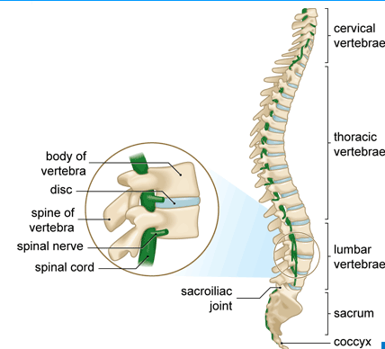
- Thoracic Spine X-ray. This is an image of the thoracic bones, which are located near the chest. There are 12 thoracic vertebrae in total.
- Cervical Spine X-ray. This is an image of the cervical bones. There are 7 cervical bones in the human body and these are located in the neck.
- Lumbosacral Spine X-ray. This X-ray includes images of the vertebrae in the lower back, known as the lumbar vertebrae. There are 5 lumbar vertebrae in the human body. Additionally, it takes pictures of the 5 fused vertebrae in the sacrum, which is located at the bottom of the spine.
- Coccyx X-ray. This is an X-ray of the sacrum and tailbone. The tailbone is also called the coccyx.
These X-rays are typically done to determine the cause of a weakness, numbness, or ongoing pain, checking for arthritis between the vertebrae or discs degeneration, checking for dislocations, fractures, infections, bone spurs, or tumors, and checking for scoliosis, congenital conditions, or changes that occur after a spinal surgery is performed.
CT scan
This type of image is also called a CAT scan. It can be thought of as a more detailed X-ray and provides doctors with a better picture of the structures under investigation. CT scans are generally used to rule out major problems but are not considered to be the most effective of all tests for back pain diagnosis.
In most cases, the patient’s back pain symptoms disappear or get better. If the back pain persists, the doctor must look for a more detailed picture of what the pain may be caused by.
-
 Asthma Diagnosis and Tests
There are several ways of making an asthma diagnosis. This article explains all the methods of asthma diagnosis....
-
9 Warning Signs & Symptoms Of Brain Tumor
Recognizing the warning signs and symptoms of a brain tumor is crucial for early detection and timely intervention....
-
Back Pain Relief: 9 Back Pain Treatment Options
There are more complex types of back pain relief treatments when it comes to this condition. To treat...
-
Osteoporosis: Symptoms, Causes, Treatments, and More
Osteoporosis is a condition affecting bone density. This article outlines the causes, risk factors, symptoms, and treatments.
-
Colon cancer: Everything you need to know
Learn about colon cancer signs and symptoms, risk factors, what to expect during a screening, and how to...
-
Osteoarthritis: Symptoms, Causes, Treatments, and More
Osteoarthritis is a degenerative joint disease that can affect joint tissues in the body. Find out its symptoms,...
-
Back Pain Symptoms: Signs and Symptoms of Back Pain
Usually, the back pain symptoms are quite obvious and include tension, stiffness and soreness. Read all the Signs...
-
What is Back Pain?
Back pain is a very common issue that almost every person experiences at a certain point in his...
-
Osteopenia: Causes, Diagnosis, Treatments, and More
Osteopenia is a loss of bone density that is not as severe as osteoporosis. Find out more about...
-
Causes of Back Pain: What Causes Back Pain?
Generally speaking, back pain can be triggered by many factors, including everyday activities. The most common causes of...


HealthNormal Editorial Team
Our qualified health and wellness editorial team, with subject matter expertise and relevant life experience, has crafted this content, upholding the highest...Read more
Brittany Heintz, MS, PhD Candidate
Brittany Heintz is a PhD Neuromechanics Candidate and Lecturer at the University of Wisconsin – Milwaukee. Her research examines how the nervous...Read more